本司产品仅用于科研,不用于临床诊断和治疗

18502057686
18502057686
Expression Host:Nicotiana benthamiana plants
Clonality:Monoclonal, recombinant
Species and Isotype: Rabbit IgG1
Description:This product is a full-length Rabbit IgG1 recombinant antibody specific to CD47 (IAP) protein. It was produced in Nicotiana
benthamiana plants via Agrobacterium tumefaciens-mediated infiltration.
Verified Applications:Western blot, ELISA, Immunocytochemistry, Flow Cytometry
Dilution Range: Western blot (1: 1 000 – 1: 2 000) ELISA (1: 1 000 – 1: 160 000)
ICC (1: 100-1: 300) Flow Cytometry (1: 300-1: 500)
Tested Species Reactivity : Human
Concentration :1 mg/ml
Form :Liquid
Storage:Short-term (up to one week): 2 – 8 °C
Long term: Aliquot and store at – 20 °C
Store immediately. Aliquot and avoid multiple freeze-thaw cycles.
Storage Buffer:0.1 M Phosphate Buffered Saline, pH 7.7. Preservative: None
Purification Notes: This product was purified using Protein A- affinity chromatography.
Purity:≥ 90% as determined by SDS-PAGE
General Notes:For Research Use only, unless otherwise indicated.
Image:
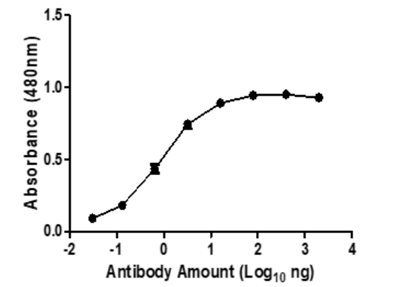
Figure 1. ELISA Dose Response curve using Rabbit Anti-CD47 Antibody from 20– 0,003 ng/uL to detect 25 ng Human CD47/Integrin Associate Protein (IAP) antigen. Experiments were performed in triplicate, with error bars representing standard deviation (SD).

Figure 2. Western blot of decreasing amounts of Human CD47 (IAP) Protein (ECD, His tag) (40 – 50 kDa smear) detected with Rabbit Anti-CD47 at 1: 1 000 and an HRP conjugated anti-Rabbit secondary antibody at 1: 10 000.

Figure 3. Immunocytochemistry: HeLa cells were plated at 200 000 cells/ well in 6-well plates on coverslips and allowed to adhere. Followed fixation and blocking, cells were incubated with 1:200 dilution of Rabbit Anti-CD47 primary antibody, and 1:300 dilution of a commercial Anti-Rabbit Alexa Fluor® 488 conjugated commercial secondary antibody. Images were taken on a Zeiss LSM780 with ELYRA PS1 platform confocal microscope (60X) at the Stellenbosch University CAF unit. Thank you to Prof Georgia Schafer (ICGEB) for kindly donating the HeLa cells and Mrs Lize Engelbrecht for her outstanding assistance.
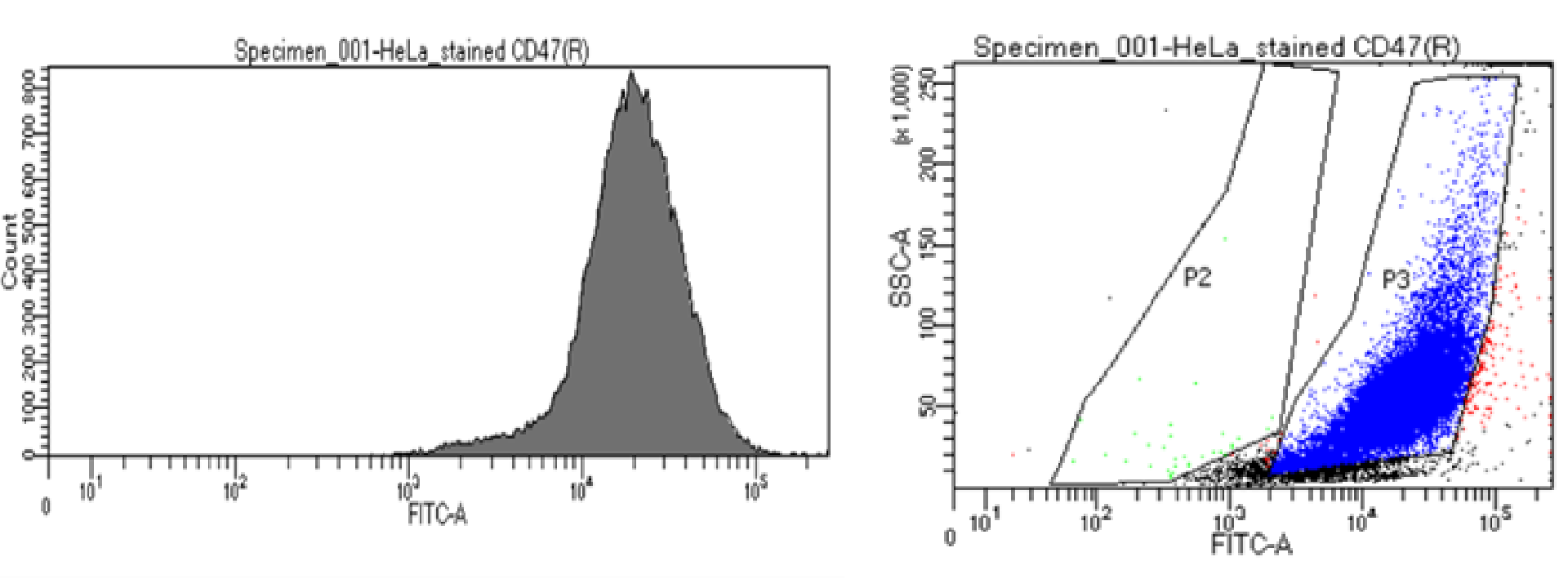
Figure 4. Flow cytometry: HeLa cells (1.3 million cells) were fixed with 4% Paraformaldehyde and washed 2x in PBS buffer. Cells were stained with 1: 400 of Rabbit Anti-CD47 (B6H12) Recombinant Antibody, followed by a commercial secondary Anti-Rabbit conjugated to Alexa Fluor® 488 at 1:400 dilution. Approximately 30 000 events were acquired on a BD FACSDiva 8.0.2 Flow cytometer. A) Dot-plot and B) Histogram of the acquired 30 000 events.
微信客服

给我留言
